Accurately measure the mass of single biomolecules in solution, in their native state, and without the need for labels.
Mass photometry is a simple, easy-to-use technology that rapidly analyzes biomolecules at the single-particle level in solution. It measures the light scattered by individual particles and uses the signal to count the particles and measure their mass with high accuracy.
Mass photometry measurements provide the mass distribution and relative concentrations of a wide range of biomolecules in a sample, accelerating a variety of studies, including:
• Sample purity and homogeneity
• Protein oligomerization
• Biomolecular interactions
• Macromolecular complex assembly

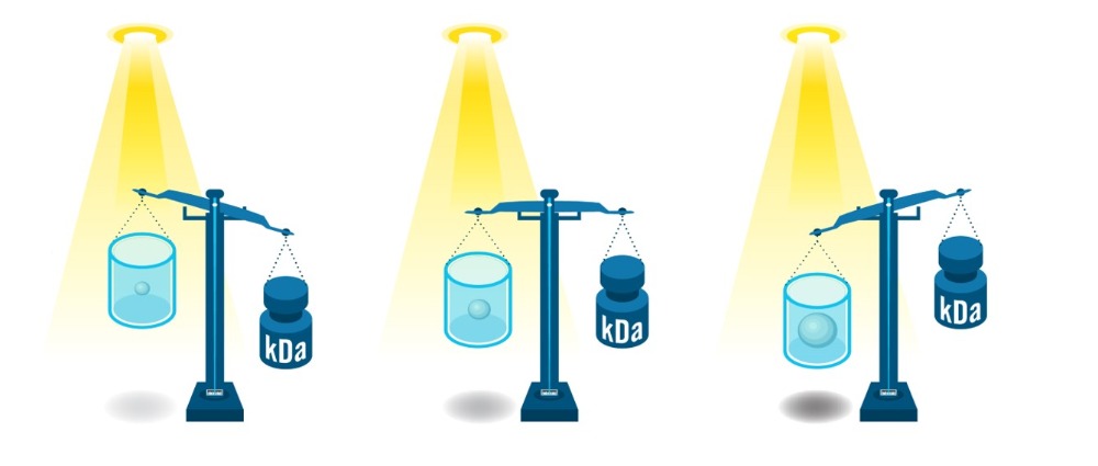
Figure1: The amount of light scattered by a particle scales linearly with the particle’s volume and refractive index. As the optical properties and density of biomolecules vary only by just a few percent, the scattering signal is directly proportional to the molecule’s mass – making it possible to weigh single molecules with light (Figure). The correlation of scattering signal with mass holds true for a variety of biomolecules (e.g. glycoproteins, nucleic acids and / or lipids), making mass photometry a universal analysis tool for biomolecules in solution.
• In solution, in a variety of buffers and compatible with membrane proteins
• Label-free, without the need to modify samples
• Single-molecule counting
• Wide mass range and high dynamic range
• Homogeneity, structural integrity and activity
• Quick, simple, minimal sample amounts
Mass photometry is a new way to analyze biomolecules, built upon the principles of interference reflection microscopy and interferometric scattering microscopy. Mass photometry enables the reliable detection of single molecules and the direct measurement of each molecule’s mass in solution.
With a wide dynamic range, mass photometry can be used to assess:
• Proteins / glycoproteins
• Nucleic Acids
• Adeno-associated viruses (AAVs)
• Antibodies
• VLPs
• Membrane proteins
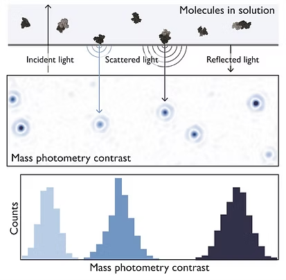
Figure 2: The principle of mass photometry. The light scattered by a molecule that has landed on a measurement surface interferes with light reflected by that surface. The interference signal scales linearly with mass.
Mass photometry enables precise quantification of the interactions between individual antibody molecules with target molecules in solution.

Our mass photometry systems support research, quality control, and manufacturing applications.
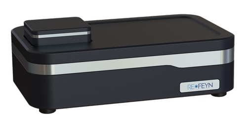
Measure molecular mass of biomolecules with speed, accuracy, and simplicity. Automated version available.
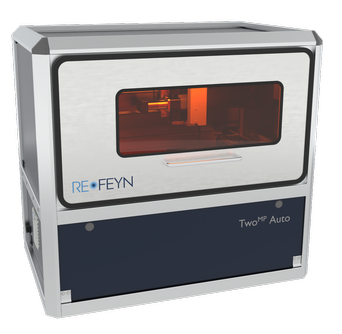
Combine the ease and efficiency of automation with the sensitivity and speed of mass photometry for rapid, accurate, multi-sample measurement.
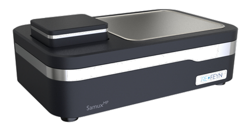
Quantitatively characterize adeno-associated viruses (AAVs) of all serotypes. An automated, high-throughput version is also available.
Mass photometry publications
– Tim Sharpe, Biozentrum, University of Basel
Explore the science of mass photometry and see how it is used to characterize protein purity, interactions, and oligomerization.
Learn the principles behind mass photometry and explore the unique benefits of this technology for biomolecule analysis.
Mass photometry on SARS-CoV-2
Basics of Mass Photometry
Formation of PROTAC ternary complexes measured with
mass photometry
Mass photometry in
denaturing conditions
Mass photometry with detergents
Characterization of protein
interaction equilibria
Quantifying heterogeneous AAV capsid loading using mass photometry
Optimizing and monitoring AAV capsid purification using mass photometry
Characterizing protein oligomerization with automated mass photometry
Quantifying protein binding affinities using mass photometry
Mass photometry analysis of low-affinity protein interactions
Mass photometry in a forced antibody degradation study
AAV titer estimation with mass photometry
Mass photometry supports membrane protein purification protocols
Accurate AAV characterization with a reliable calibrant for mass photometry
Streamlining AAV analytics in gene therapy manufacturing workflows
Antibody aggregation analysis: Mass photometry vs. SEC-UV
Mass photometry of nanocages
Mass photometry analysis of GPCRs in copolymer nanodiscs
Quantifying adenovirus packaging with macro mass photometry
Streamline antibody stability analysis with mass photometry
Measuring AAV genome length with mass photometry
Measuring protein-nucleic acid interactions with mass photometry
Upstream AAV characterization with mass photometry: Application note and Protocol